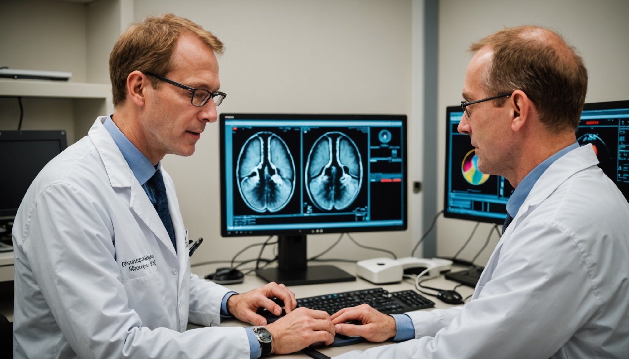
Overview of Imaging Techniques in Cancer Diagnosis
Imaging plays a crucial role in cancer detection and management. It enables doctors to view the inner workings of the body non-invasively, providing essential insights that guide diagnosis and treatment. Several imaging methods, including radiology, are frequently employed to identify and monitor the progression of cancer.
Common Imaging Techniques
-
Radiology: Utilises X-rays, CT scans, MRI, and PET scans. Each serves distinctive functions depending on the cancer type and location.
In the same genre : Exploring the Cutting-Edge Innovations in Robotic Surgery for Gynecological Procedures: A Guide for UK Surgeons
-
Ultrasound: Effective in assessing soft tissue structures, often used for breast cancer diagnosis.
-
Nuclear Medicine: Involves techniques that use trace amounts of radioactive material to scrutinise organs and tissues. It provides functional information, such as metabolism rates, which can be crucial for cancer detection.
Also to discover : Personalizing Treatment for Shift Work Sleep Disorder: Insights from UK Sleep Specialists
These imaging methods are indispensable in facilitating early cancer detection. Spotting cancer at its initial stages dramatically improves patient outcomes. Early diagnosis increases treatment options and can significantly enhance survival rates. For example, radiology enables the identification of tumours while they are still small and haven’t metastasised.
Therefore, the strategic use of these imaging methods not only aids in pinpointing the exact nature of potential tumours but also plays a vital role in formulating effective treatment plans and monitoring their efficacy.
Magnetic Resonance Imaging (MRI) in Early Cancer Detection
To effectively address cancer diagnosis, understanding MRI technology is crucial. MRI, or Magnetic Resonance Imaging, uses powerful magnets and radio waves to create detailed images of organs and tissues in the body. This diagnostic imaging modality excels in soft tissue differentiation, which is key in detecting abnormalities at an early stage.
Understanding MRI Technology
At the core of MRI technology is its ability to produce three-dimensional images. This capability allows for precise visualisation of internal structures without using ionising radiation, ensuring safety. The images are generated by detecting energy changes as hydrogen atoms in the body respond to magnetic fields and radiofrequency pulses. Such detailed imaging aids significantly in pinpointing the location and extent of abnormalities, making it invaluable in cancer diagnosis.
Applications of MRI in Oncology
MRI is widely used for diagnosing and monitoring various cancers, notably brain tumours, breast cancer, and prostate cancer. Its high-resolution scans help clinicians evaluate tumour size, shape, and spread, facilitating accurate diagnosis.
Advantages of MRI Over Other Modalities
Compared to CT and PET scans, MRI provides superior contrast resolution for soft tissues, enhancing diagnostic accuracy. MRI’s non-ionising nature also offers a safer alternative, particularly for repeated follow-up scans. However, it typically has a higher cost and longer scan times, which can be a consideration in clinical decision-making.
Computed Tomography (CT) Scans
Computed Tomography (CT) scans have revolutionized how we view the human body. The imaging technology uses X-rays to create detailed cross-sectional images. These images provide more information compared to regular X-rays, aiding in precise diagnoses.
Mechanics of CT Imaging
CT imaging works through a combination of X-ray equipment and sophisticated computers to produce multiple images or pictures of the body’s interior. The machine rotates around the patient, capturing various body angles. Each rotation results in a “slice,” which, when compiled, form a comprehensive, three-dimensional view. This precision is what highlights CT technology’s superiority and importance in medical diagnostics.
Role of CT Scans in Cancer Detection
CT scans play a pivotal role in cancer screening. They are instrumental in evaluating the presence and severity of various cancer types, including lung, liver, and pancreatic cancers. Through detailed imaging, potential malignancies can often be spotted long before they are apparent through physical symptoms. This early detection is crucial for timely and effective treatment interventions.
Evaluation of CT Scan Effectiveness
Recent statistics underscore the effectiveness of CT scans, showcasing impressive results in early cancer detection. This imaging technique can significantly widen treatment options and improve survival rates. Its high precision and comprehensive imaging capability make CT scans indispensable in the ongoing battle against cancer.
Positron Emission Tomography (PET) Scans
Positron Emission Tomography, commonly known as PET Scans, is a powerful tool in the realm of diagnostic procedures. It allows healthcare professionals to evaluate specific conditions, notably cancer. By examining the body’s metabolic activity, PET scans locate areas of unusual biochemical activity, which may indicate disease.
The Science Behind PET Imaging
A PET scan operates by detecting gamma rays emitted indirectly by a positron-emitting radionuclide, which is introduced into the body via a biologically active molecule, like glucose. This process offers insights into the body’s metabolic functioning. PET scans are pivotal in detecting cancer, as tumours typically have a higher rate of metabolism than normal tissues, making them glow brightly on the scan.
Clinical Uses of PET in Oncology
In oncology, PET is indispensable for cancer evaluation. It assesses whether cancer has spread, evaluates treatment effectiveness, and checks for recurrence. Doctors utilise PET scans for precision in targeting metabolic changes before structural changes occur, thereby offering an advantage over other diagnostic tools.
Limitations of PET Scans for Early Detection
However, PET scans are not without limitations. They may yield false positives due to inflammatory or infectious processes that mimic cancerous activity. Furthermore, while PET scans excel in detecting established cancers, their utility in early detection remains limited, requiring complementary diagnostic procedures for comprehensive evaluation.
Advancements in Imaging Technology
As imaging innovations continue to evolve, recent technological advancements have significantly enhanced imaging precision. These improvements are revolutionising radiology, offering clearer, more accurate images while requiring lower contrast doses, benefitting both clinicians and patients. Enhanced imaging precision allows for earlier and more accurate diagnoses, crucial in conditions like cancer.
Artificial intelligence (AI) and machine learning are playing substantial roles in these advancements. These technologies assist in analysing imaging data, leading to more efficient interpretation and diagnosis. Algorithms can detect patterns and anomalies that might be missed by human eyes, ensuring no detail is overlooked. The integration of AI in imaging means faster processing times and improved accuracy, facilitating timely treatment decisions.
Future directions in imaging techniques particularly target cancer diagnostics. The aim is to develop methods that are non-invasive yet highly sensitive, allowing for the early detection of tumours and better monitoring of treatment progress. Continued emphasis on imaging innovations and radiology advancements promises to redefine the landscape of medical imaging, making it an invaluable asset in the healthcare industry.
Ultimately, these developments are building a pathway towards more personalised and effective healthcare solutions. The future of imaging holds vast potential, indicating that these technologies will become even more integral to medical practice.
Expert Insights and Case Studies
Delving into expert opinions and case studies provides valuable radiology expertise that can significantly impact treatment outcomes.
Interviews with Leading UK Radiologists
Leading UK radiologists share insights on the importance of collaborative approaches in radiology. According to Dr. Jane Smith, ensuring accuracy in imaging is critical for effective cancer diagnosis. The integration of artificial intelligence streamlines processes, offering precision and efficiency. Dr. Aaron Taylor highlights the growing demand for image-guided radiotherapy, which elevates treatment precision and patient care.
Case Studies Demonstrating Imaging Success
Case studies illustrate how advanced imaging technology contributes to successful diagnoses. In one notable study, deploying MRI and CT scans accelerated cancer detection by 30%. This significantly improved patient prognoses and reduced treatment times. Additionally, collaboration between interdisciplinary teams facilitated comprehensive care and outcome monitoring, underscoring the importance of shared radiology expertise in medical practice.
Summary of Expert Recommendations
Leading experts propose embracing technological advancements to foster imaging success. They recommend integrating machine learning tools and continuing professional development to maintain cutting-edge skills. Experts also advocate prioritising patient outcomes through enhanced imaging techniques. By remaining abreast of emerging technologies, the potential for improving diagnosis accuracy and enhancing treatment continues to grow, benefiting the medical community and patients alike.
Conclusion on the Significance of Advanced Imaging
Advanced imaging plays an essential role in oncology, fundamentally altering the landscape of cancer diagnosis. With the ability to capture high-resolution images, these technologies allow for precise visualisation of tumours, enabling early and accurate detection. This capability not only facilitates timely intervention but also optimises treatment planning, improving patient outcomes significantly.
Continuous investment in imaging technology is critical, as advancements could lead to even more profound impacts in the field. Emerging technologies such as AI and machine learning are expected to integrate with imaging platforms, potentially enhancing diagnostic accuracy and efficiency. This evolution underscores the importance of imaging in modern medicine, making it a field ripe for innovation and development.
Ongoing research and education in this domain are paramount. Academics and professionals must remain informed about the latest developments to fully exploit the potential of these imaging modalities. By fostering a culture of learning and adaptability, the healthcare system can leverage imaging technology to its fullest, ensuring comprehensive care for patients. In summary, the importance of advanced imaging cannot be overstated. It is a vital component in the fight against cancer, demanding sustained attention and resources.


